Anatomy of the thyroid gland
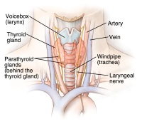 |
| Thyroid Gland - Click to Enlarge |
The thyroid gland is located in the front of the neck (just above the breastbone and below the Adam's apple). The small, two-inch gland consists of two lobes, one on each side of the windpipe, connected by a small bridge of thyroid tissue called the isthmus.
The thyroid tissue is made up of two types of cells: follicular cells and parafollicular cells. Most of the thyroid tissue consists of the follicular cells, which secrete the iodine-containing thyroid hormones. They consist of thyroxine (T4) and triiodothyronine (T3). The parafollicular cells (also called C cells) secrete the hormone calcitonin. Calcitonin is a weak hormone that helps in the regulation of calcium in some animals. In humans, calcitonin has only a minor role in calcium regulation. When the thyroid is completely removed because of cancer or disease, the removal of these C cells produces no problems with calcium regulation. The follicular cells require an adequate supply of iodine in order to produce the thyroid hormones.
Functions of the thyroid gland
The thyroid plays an important role in regulating the body's metabolism. Virtually every tissue in the body is affected or regulated by thyroid hormone. It regulates our metabolism, brain development and function, skin, hair, eyes, heart, and intestine function. The thyroid hormones enter into tissues and regulate how those tissues produce or do not produce certain proteins. The thyroid function is controlled by the pituitary, which sits at the base of the brain. The pituitary is controlled by a region in the brain called the hypothalamus.